Neurorehabilitation » Traumatic brain injury » TBI: General Information, Articles » Classification of the head injuries
Classification of the head injuries
Concussion
Concussion is an instantaneous failure of the brain to perform its functions, the most common form of traumatic brain injury is accompanied by a brief loss of consciousness. Once the patients regains consciousness, they complain of headache, dizziness, nausea and often vomiting, tinnitus, sweating and sleep disorders. The vital functions do not suffer significant deviations. The general condition usually improves during the first, rarely the second day. If any symptoms are found, the patient should immediately undergo a medical examination to exclude a concussion. This TBI is treatable.
Focal direct injury
Any force that has any effect on the head or leads to a skull fracture causes severe brain injuries. Thus, the shock wave spreading from the area of blow through the brain and to the opposite pole with rapid pressure drops in the areas of blow and countercoup leads to a number of serious complications. In the case of a fracture, the fragments of the skull that have penetrated into the brain can cause tissue damage. A direct focal injury occurs when the head (skull) strikes, for example, against the floor when a person falls or when it strikes against the steering wheel as a result of a car accident.
Although as a result of such injuries skull remains intact, has no cracks and fractures, the shock wave leads to a concussion of the brain itself. When a person after a stroke sharply stops the movement of the head, the brain still continues to move hitting the walls of the skull, which causes a bruise of the brain and bleeding (hemorrhage). This is due to the hydrodynamic effect of the liquor wave. In such cases, the injuries occur closer to the part of the brain where the blow occurred. It is most commonly frontal and temporal lobes. In cases of blunt head injury, the brain damage can be directly opposite to the site of the blow. Such an injury is called damage caused by an anti-blow. This usually happens when the head hits against a stationary object, for example, against a windscreen.
Diffuse indirect injury
In addition to the focal direct injury, the research has found another type of TBI. This phenomenon is called shaken baby syndrome, as well as a syndrome of a shaken child, a syndrome of a child's traumatic shaking. Shaking significantly stretches and damages sensitive nerve cells, which sometimes leads to very significant damages or even death. When adults have severe injuries of the back, neck, the force of the blow also has a detrimental effect on the brain, thereby causing irreversible brain damage.
Diffuse axonal injury
Diffuse axonal injury (DAI) is a common type of TBI, in which a sharp acceleration or braking of the head, for example, at the time of an accident, leads to axonal tension and rupture. The brain consists of billions of nerve cells located in a gray matter, which are connected by long nerve fibers. These fibers are called axons and make up a white matter. Other less common causes of DAI can be falls, blows during a fight or beating.
The diffuse axonal injury results in the detection of microscopic small focal hemorrhages in the calloused body, semi-oval center and upper parts of the brainstem. Clinically, it manifests itself as a prolonged coma, which in most cases changes into a vegetative state. The latter is characterized by a lack of cortical activity and lasts for months or years.
Although injuries of this kind usually occurs as a result of severe damage to the cervical spine that leads to a coma. Recent studies have shown that diffuse axonal injuries also develop, albeit to a lesser extent, due to a brief loss of consciousness. Since such injuries cause microscopic damage, it is impossible to capture them on CT or MRI.

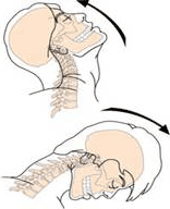
Side view, movement of the head and neck during a cervical injury.
The damage to the brain also occurs as a result of the injury, which occurred at high speed. These can be a fall from a high altitude, high speed during a car accident. Either deformation or stretching of the brain tissue occurs, which leads to tissue rupture. Such an injury causes damage to the brain due to "pulses" or "waves" passing through the brain at extremely high speeds. The damage occurs as a result of a dramatic change in density within individual brain cells and instantaneous contractions lead to damage of the internal structures of the brain cells.
Many similar injuries were found in veterans who returned from the war. They emerged as a result of the explosion or explosive charges near them. Pressure or pulses from the explosion passed through the human body. It passing through the brain caused damage to the cells. Although many of these soldiers did not have any visible symptoms, complaints, there are no guarantees that in the future this will not lead to the development of serious brain disorders.
Hypoxia or poisoning from toxic substances
The human brain is more susceptible to injuries resulting in a lack of oxygen in the body (hypoxia) than any other part of the body. Hypoxia can occur in combination with other injuries (infarction) or in any other situation, when the breathing process is disrupted and the supply of oxygen to the body stops. The complications arising from hypoxia often have a negative effect on the hippocampus.
The hippocampus is part of the limbic system of the brain (olfactory brain). It participates in the mechanisms and the formation of emotions, the consolidation of memory, i.e. the transition of short-term memory to long-term memory. The exposure to toxic chemicals (lead, toluene, carbon monoxide) can also cause brain damage. The degree of damage depends on the level of exposure to the substance and its duration (dose).
Accompanying Brain Injuries
In addition to direct neurological brain injuries, there are a number of brain injuries that do not have a neurological nature.
Cerebral edema is a life-threatening pathological process manifested by excessive accumulation of fluid in the cells of the brain or spinal cord and intercellular space, an increase in brain volume and intracranial hypertension. Cerebral edema becomes dangerous when the tumor provokes an increase in intracranial pressure, which prevents the ingress of blood into the cranial cavity to deliver glucose and oxygen to the brain. When a person suffers from constant intracranial pressure, they are prescribed treatment, which helps to normalize it. In more complex cases, one needs surgical intervention. A hole is made in the skull to create an outflow of a part of the cerebrospinal fluid, which leads to an increase in intracranial pressure.
Hematoma is a type of bruising, limited blood accumulation with closed and open tissue damage with vascular rupture. In this case, a cavity containing liquid or coagulated blood is formed. Computed tomography can detect such bruises. The cerebral hemorrhage caused after an injury can only show a few days after the patient's discharge. The dura mater covers the entire brain and spinal cord. The blood clots formed outside the meninx, between the inner surface of the skull and the outer layer of the dura mater (epidural space), are called epidural hematoma. Also, there is a subdural hematoma. This is the accumulation of blood in the space between the inner leaf of the dura mater and the arachnoid matter. The arachnoid matter is one of the three membranes that cover the brain and spinal cord. It is located between the two other meninges: the most superficial dura mater and the deepest pia mater separated from the latter by the subarachnoid space. This cavity is filled with 120-140 ml of cerebrospinal fluid. The cerebrospinal fluid is a liquid that constantly circulates in the ventricles of the brain, the liquor conducting pathways, the subarachnoid space of the brain and spinal cord. When blood gets into the cerebrospinal fluid, it results in a hemorrhage.
Hydrocephalus and hygroma. Hydrocephalus or water in the brain, is a disease characterized by an excessive accumulation of cerebrospinal fluid in the ventricular system of the brain due to its difficulty moving from the area of secretion (the ventricles of the brain) to the area of absorption into the circulatory system (subarachnoid space) or due to the impaired absorption. If the blood somehow gets into the cerebrospinal fluid and blocks the areas of absorption, the cerebrospinal fluid returns back to the ventricles increasing their size. If the pressure inside the ventricles is greatly increased and can lead to a risk of brain damage, the patient is injected with special tubes into the ventricles, which help reduce pressure. Hygroma is the accumulation of cerebrospinal fluid in the subdural space. If the swelling in the hygroma exerts a strong pressure on the brain, the person requires surgical intervention.
 |
 |
 |
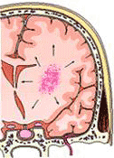
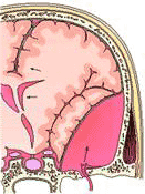
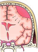
| Intracerebral hemorrhage. Central large dark areas show a hemorrhage. | Epidural hematoma. The dark area in the lower right region indicates a hematoma. Broken blood vessels and displacement of median structures were recorded. | Subdural hematoma. Hematoma is shown by the dark areas in the upper right corner. |
Here you can also read other articles on this topic:
To receive professional advice on the treatment of traumatic brain injuries in GermanyPlease cll us: +49 228 972 723 72
or write an Email here


